
When someone develops a respiratory infection, an important part of the body’s defense takes place inside the lungs.
But understanding exactly how those defenses work has been a persistent challenge.
Researchers can study immune cells in blood samples. They can examine lung tissue. They can also conduct experiments in animals.
What has been much harder is studying living human lung tissue while preserving the complex network of immune cells that normally helps protect it.
Now, scientists at Stanford University School of Medicine have demonstrated a promising new approach.
In a study published in Nature on October 7, 2026, researchers developed laboratory models that allowed them to observe how human lung tissue responds to viral infection.
The achievement could open new possibilities for respiratory disease research.
Keeping the Lungs’ Natural Defenses Together
The researchers worked with human lung tissue obtained from more than 220 patient volunteers.
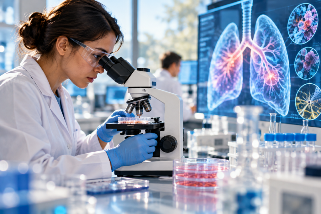
Using a specialized laboratory system, they maintained small fragments of lung tissue for extended periods while preserving several types of immune cells.
These laboratory models are called organoids.
Unlike simpler cell cultures, the new models preserve elements of the natural relationships between lung tissue and its resident immune system.
That matters because the lungs are not passive organs waiting for immune cells to arrive from elsewhere in the body.
They contain immune cells that can recognize threats and respond locally.
The researchers were particularly interested in T cells, which play an important role in identifying and responding to infections.
Their laboratory models preserved these cells and allowed scientists to investigate how they reacted when the tissue encountered a virus.
What Happened When Researchers Introduced a Virus?
The team exposed the laboratory lung models to SARS-CoV-2, the virus responsible for COVID-19.
They observed several immune responses, including inflammation and the activation of T cells that specifically recognized the virus.
The researchers then carried out another experiment.
Before exposing the lung models to the virus, they stimulated their immune cells using viral protein fragments.
This was designed to investigate whether the lung’s local immune defenses could be prepared to respond more effectively.
The results were encouraging.
The stimulated immune cells showed a virus-specific response, and subsequent viral infection in the models was reduced.
The findings suggest that immune cells already present in lung tissue may play an important protective role.
They also provide researchers with a new way to investigate whether future vaccination strategies could strengthen these local defenses.
Why This Could Matter for Future Vaccines
Most people are familiar with vaccines as a way of preparing the immune system to recognize an infectious threat.
But researchers are increasingly interested in understanding how protection works at the specific locations where infections begin.
For respiratory viruses, that includes the tissues lining the airways and lungs.
A better understanding of these local defenses could eventually help scientists investigate new approaches to vaccination.
For example, researchers may be able to examine whether certain vaccine strategies produce stronger protective immune responses directly within respiratory tissue.
The Stanford study does not establish that such a vaccine will work in humans.
Instead, it provides a laboratory system for investigating questions that have previously been difficult to answer.
That distinction is important.
Medical advances often begin with better research tools, long before a treatment becomes available to patients.
A More Human Way to Study Human Disease
Another promising aspect of the research is its potential to improve how respiratory diseases are studied.
Animal models remain important in biomedical science, but immune responses observed in animals do not always reproduce what happens in people.
Laboratory models built from human tissue may help researchers understand some of these differences.
The National Institutes of Health also noted that this approach could potentially replace certain animal-model experiments, although it has not been demonstrated as a comprehensive alternative.
The researchers believe similar methods may eventually help them investigate other respiratory conditions, including asthma, chronic obstructive pulmonary disease and bacterial infections.
These applications remain possibilities for future research rather than established medical benefits.
An Important Step, Not Yet a New Treatment
The findings deserve attention, but they also require perspective.
The research was conducted using laboratory-maintained human lung tissue.
It did not demonstrate that stimulating lung immune cells protects patients from infection.
It did not establish the effectiveness of a new vaccine.
And it does not change existing medical recommendations.
Further research will be needed to understand how the findings translate to living people and whether they can contribute to safe, effective preventive treatments.
Nevertheless, the study represents a meaningful development: scientists can now investigate important aspects of human lung immunity in a model that preserves more of the tissue’s natural complexity.
That could help answer questions that were previously difficult to study.
NewsPositive Takeaway
Sometimes a promising medical breakthrough is not a new medicine or a treatment that immediately changes people’s lives.
Sometimes it is a better way to understand how the human body already protects itself.
The Stanford researchers have created a tool that could help scientists study respiratory infections more realistically and investigate new ways of strengthening the lungs’ natural defenses.
There is still much to learn.
But each improvement in our ability to understand human biology creates opportunities for future discoveries.
And that is something worth feeling hopeful about.
Good things are happening. We’re here to find them.
Sources
- Nature — Human lung organoid modelling of tissue-resident antiviral T cell responses
- National Institutes of Health — Human lung organoids with a built-in immune system could transform respiratory disease research
Editorial note: The organoid system and immune-response findings are established laboratory results. Potential benefits for future vaccines, respiratory treatments, or reduced reliance on animal models remain future research applications and are not yet clinical outcomes.